
If you’ve been following a low histamine diet for eczema and found yourself still flaring three weeks in, here’s what I need you to understand: histamine didn’t cause your eczema. It responded to it. Every protocol that starts with “eliminate fermented foods and leftovers” is treating a symptom that is at least two or three layers removed from what’s actually driving the inflammation.
In my functional medicine practice, I have never met a patient whose eczema was caused by histamine. What I’ve consistently seen is elevated histamine as a downstream signal — the visible end of an inflammatory cascade that began months or years earlier in hormone metabolism, liver detoxification pathways, and microbial terrain. The low histamine diet offers temporary relief because it reduces the load on a system already overwhelmed. But the system itself remains broken. That is why elimination helps but never heals.
This is what I test for instead. And the clinical pattern — across hormone panels, toxin panels, and micronutrient testing — tells a very specific story.

Histamine Is Downstream, Not the Diagnosis
Histamine is a biogenic amine produced by mast cells, basophils, and certain bacteria in the gut. Your body makes it, bacteria make it, and you consume it in food. Under normal circumstances, two enzymes keep it in check: diamine oxidase (DAO), which breaks down dietary histamine in the gut lumen, and histamine N-methyltransferase (HNMT), which inactivates histamine inside cells systemically.
When these systems fail, histamine accumulates and symptoms follow — the itch, flush, hive, and swelling that look like allergy but test negative for it. The standard clinical response is a low histamine diet: remove fermented foods, aged cheeses, leftover meats, alcohol, and high-histamine produce. This reduces histamine intake. And it usually helps to some degree. For a while.
Here is the clinical problem: DAO deficiency is itself downstream. DAO production is compromised by gut mucosal damage, intestinal permeability, dysbiosis, and nutritional deficiencies in the cofactors it requires — specifically vitamin B6, copper, and vitamin C. Every one of those — the damaged gut lining, the dysbiosis, the micronutrient insufficiency — is caused by something else. Eliminating fermented foods doesn’t rebuild the gut mucosa. It doesn’t rebalance the microbiome. It doesn’t address the hormonal or toxic burden that inflamed the gut wall in the first place.
The question I ask every patient with eczema who reports that histamine-restricted eating helps somewhat but never resolves the problem is not “how do we lower histamine?” It is “what is driving the immune activation that keeps triggering histamine release?” Those are completely different clinical questions, and they lead to completely different interventions.
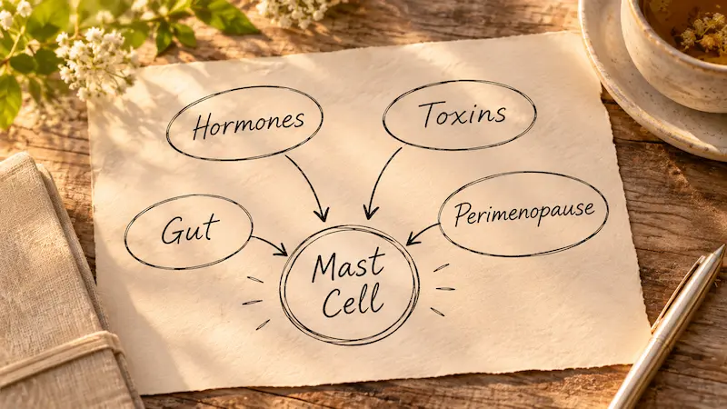
The Four Drivers Keeping Your Mast Cells Primed
In the eczema patients I see in practice, there are four distinct drivers — often present simultaneously — that maintain mast cells in a state of chronic sensitization. Sensitized mast cells release histamine at a lower threshold than normal, which is why these patients react to foods, environmental exposures, and stress responses that shouldn’t be significant triggers.
Driver 1 — Hormone dysregulation through poor estrogen clearance
Specifically, an imbalance in phase 1 estrogen metabolism that routes estrogens toward genotoxic and proliferative pathways rather than the protective 2-hydroxylation pathway. I’ll explain the clinical evidence for this in the next three sections because it’s the driver most consistently underidentified in dermatology and allergology settings.
Driver 2 — Toxic overload and redox imbalance
Accumulated environmental toxins — primarily mycotoxins from food sources — that exceed the body’s biotransformation capacity, generate oxidative stress, and directly activate mast cell degranulation through TLR4/NF-κB inflammatory signaling.
Driver 3 — Dysbiosis and Th2 immune skew
An altered gut microbiome that tips the immune system toward the Th2 response pattern specifically associated with atopic dermatitis. Th2 dominance heightens IgE-mediated and mast-cell-mediated reactivity across all tissue.
Driver 4 — Perimenopause convergence
Not a separate driver but the clinical moment when drivers 1 through 3 collide with the hormonal shifts of midlife. This is when many of my patients have their first significant eczema onset, or when long-standing mild eczema becomes severe and unmanageable. None of these are addressed by dietary histamine restriction alone.
The Estrogen-Histamine Axis — What Conventional Allergology Misses
Mast cells are estrogen-sensitive. They carry estrogen receptors, and elevated estradiol directly triggers mast cell degranulation and histamine release. This means that any state of estrogen excess or poor estrogen clearance creates a continuous background stimulus for histamine production that has nothing to do with what you ate.
The source of that estrogen excess is rarely a single problem. In my functional medicine practice, I see it emerge from two converging mechanisms.
The first is impaired hepatic clearance. Estrogen is processed in two phases of liver detoxification. Phase 1, primarily through the CYP3A4 enzyme, converts parent estrogens into hydroxylated metabolites. Phase 2 then conjugates these metabolites — through methylation, glucuronidation, and sulfation — for safe elimination. When phase 2 capacity is impaired by nutritional deficiencies, genetic variants on methylation enzymes, or oxidative stress, metabolites accumulate between the two phases. This is estrogen burden. And it feeds the mast cell activation loop continuously.
The second mechanism involves the aromatase enzyme. Aromatase converts testosterone into estradiol and estrone, and it is expressed in adipose tissue. This means adipose tissue is not metabolically passive — it is actively producing estrogen. The “extra weight that just won’t move” that so many of my eczema patients describe is not simply a symptom. It is an estrogen factory. As adipose mass increases, aromatase activity increases proportionally, and the estrogen burden compounds.
The relationship between weight gain that resists conventional intervention and the onset or worsening of eczema is not coincidental. I see it clinically, repeatedly. The low testosterone that often accompanies this pattern — frequently passed over because it falls “within normal limits” on standard labs — is the other side of the same coin: testosterone being preferentially routed into estrogens via aromatase rather than supporting its own function.
Reading the Pattern — A Clinical Teaching Case
The de-identified Vibrant Wellness 24-hour urinary hormone panel included with this post shows the metabolite pattern I see often in eczema patients with the hormonal driver. I am sharing it because the visual makes what I’ve described clinically concrete in a way that text alone cannot.
The panel shows estradiol significantly elevated above the reference range — more than twice the upper limit — alongside low testosterone and low estriol. That combination tells a specific story: active estrogen excess, with testosterone being preferentially converted through aromatase rather than supporting its own function, and poor conversion to estriol, the least biologically active of the three estrogens.
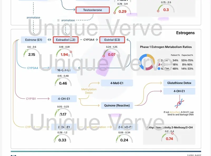
But the more important finding is in the Phase 1 Estrogen Metabolism Ratios. Before looking at how estrogen is being metabolized, note what it’s being metabolized from: estradiol at 1.94 against a reference range of 0.18–0.81 — more than twice the upper limit. The metabolite pattern below is downstream of that burden.
- 2-hydroxylation (2-OH): The protective pathway. 2-OH metabolites are weak, non-carcinogenic, and clear easily. This patient’s 2-OH-E1 was 0.33 — sitting at the low end of a reference range of 0.28–1.2. The protective pathway was barely active, producing the minimum output needed to stay technically within range while contributing almost nothing to meaningful estrogen clearance.
- 4-hydroxylation (4-OH): The genotoxic pathway. 4-OH-E1 was 0.17 against a reference range of 0.05–0.23 — within range, but in the upper third. When the 2-OH pathway is underperforming and estrogen burden is this high, even a mid-range 4-OH value represents a disproportionate genotoxic load relative to available clearance capacity.
- 16-hydroxylation (16-OH): The proliferative/estrogenic pathway. 16-OH was 0.46 against a reference range of 0.15–0.48 — right at the upper ceiling of normal. 16-OH metabolites retain estrogenic activity and directly sensitize estrogen-responsive tissue, including mast cells. The pathway was running at full capacity with no headroom.
What this means clinically: very little estrogen was going through the protective route. Most was going through pathways that either directly damage tissue or maintain a state of prolonged estrogenic stimulation. The mast cells — continuously signaled by elevated estradiol and elevated 16-OH metabolites — have every biochemical reason to stay primed for histamine release. And they do.
The 4-OH Cascade — Why This Metabolite Threatens More Than Your Skin
The 4-OH pathway deserves deeper explanation because what happens inside it is not just about skin inflammation. It is a process of ongoing cellular damage, and understanding it explains why long-standing histamine-driven eczema almost always signals a broader systemic problem.
Think of estrogen not as a single substance but as a raw material your body has to process and neutralize. The processing happens in stages. At one particular stage, estrogen gets converted into a form that is significantly more reactive than the original. What happens next is what matters for your health beyond eczema.
Step 1 — Estrogen gets converted into a more reactive form
An enzyme in your liver takes circulating estrogens and converts them into a chemically more active version — one that your body now has to neutralize quickly, before it causes damage. If neutralization is slow or impaired, this reactive form accumulates.
Step 2 — That reactive form becomes something that damages cells
The activated estrogen undergoes another conversion, producing a compound called a quinone. A quinone is not just reactive — it actively seeks out other molecules to bond with. Think of it as a chemically “grabby” molecule that latches onto whatever biological structure is nearby. At this stage, the estrogen metabolite has stopped behaving like a hormone byproduct and started behaving like a cellular irritant.
Step 3 — It attaches to your DNA
The most available target for a quinone is DNA — specifically the chemical bases that make up your genetic code. The quinone bonds to these bases and pulls them out of the DNA strand, leaving gaps behind. Your cells try to repair those gaps, but repair is imperfect. The errors left behind are mutations — permanent changes in the genetic sequence of that cell. This is not theoretical cancer biology. It is a documented mechanism that runs continuously when elevated 4-OH estrogen metabolites are not being adequately cleared.
Step 4 — The process generates ongoing oxidative stress
Here is what makes this more than a one-time event: the quinone doesn’t cause damage and disappear. It cycles back and forth between two forms, and each cycle generates free radicals — unstable molecules that damage everything they contact: cell membranes, proteins, more DNA. This is what oxidative stress means at the cellular level. It is not acute inflammation triggered by a food or allergen. It is a continuous background process driven by the estrogen metabolite itself, running independently of anything you eat or avoid.
Step 5 — The free radical damage keeps mast cells permanently activated
Sustained oxidative stress triggers one of the body’s most powerful inflammatory switches. When that switch is activated, it turns on the production of inflammatory messengers — the same ones that keep mast cells in a sensitized, hair-trigger state. Sensitized mast cells release histamine at a lower threshold than normal. They react to foods, smells, stress, and temperature changes that shouldn’t trigger a significant response in a healthy immune system.
The mast cell is not the problem. It is a downstream consequence of an inflammatory signal that began with an estrogen metabolite that wasn’t cleared.
This is why the low histamine diet addresses the last link in the chain rather than the chain itself. And it is why the eczema, in patients with this hormonal profile, is often the most visible sign of a broader process simultaneously affecting cellular integrity, immune regulation, and long-term disease risk.
The two detox exits — and why they fail
The body has two mechanisms to neutralize quinones before they reach DNA. When both are compromised, the cellular damage cycle runs continuously.
The first exit is COMT-mediated methylation. The enzyme catechol-O-methyltransferase (COMT) methylates 4-OH-E1 to the inert 4-MeO-E1, which clears safely. This requires SAM (S-adenosylmethionine) as the methyl donor, which depends on adequate B12, folate, and B6. Genetic variants on the COMT gene reduce baseline clearance capacity. And — critically — cortisol competes with estrogen metabolites for the same COMT enzyme. Every stress response and every cup of coffee that elevates cortisol reduces the capacity to clear 4-OH catechol estrogens through the methylation exit.
The second exit is glutathione conjugation, which neutralizes quinones before they reach DNA. This requires adequate reduced glutathione — a resource chronically depleted in patients with high toxic burden, impaired phase 2 function, or genetic variants on glutathione transferase genes (GSTM1, GSTT1). When glutathione is insufficient, the quinone pathway cannot be quenched.
When both exits are compromised simultaneously — which is the common clinical picture in eczema patients with this hormonal profile — the quinone reaches the DNA. The eczema is not the worst thing happening in this patient’s body. It is the most visible.
The Mycotoxin Problem Hiding in “Healthy” Food
One of the most consistent findings in my eczema patients is the presence of mycotoxins — fungal metabolites that contaminate grain crops during storage and processing. The pattern is so consistent that it no longer surprises me: the patient eating what they believe is a clean, anti-inflammatory diet has often been quietly loading mycotoxin burden through the very foods they considered healthy.
Mycotoxins act directly on the immune system through NF-κB signaling — the same inflammatory pathway downstream of the 4-OH quinone cascade described above — and have been shown to promote sustained mast cell hyperactivation rather than the normal episodic mast cell response. This is not allergic mast cell activation. It is toxic mast cell activation, and it explains why antihistamines and low-histamine diets provide only partial relief: they address the histamine output but not the mast cell activation signal upstream of it.
The grains most implicated are conventional wheat, corn, and oats — eaten in forms that appear genuinely health-conscious: “healthy whole grains,” gluten-free products made with corn starch and rice flour, oat-based products marketed for gut health. The mycotoxin contamination occurs during storage in conditions that allow fungal growth, and many of the toxins — particularly ochratoxin A, aflatoxin, and deoxynivalenol — survive cooking and processing. Organic certification does not eliminate mycotoxin risk. Properly dried and stored grains reduce it. But grains are categorically a mycotoxin vector in a way that most dietary guidance doesn’t acknowledge.
When I see eczema patients who assumed their grains were healthy, this is what I explain: clean eating and high mycotoxin load are not mutually exclusive. The “bucket overflow” model is useful here — the total toxic burden your detoxification system is managing at any given time determines your inflammatory threshold. Add mycotoxins from food to poor estrogen clearance and an already-inflamed gut, and the bucket overflows at a much lower provocation.
Why Coffee Makes Eczema Worse (And It Has Nothing to Do With DAO)
You’ve probably read that coffee inhibits DAO and raises histamine levels. That mechanism exists. But in my patients, the more clinically significant reason is what coffee does to the estrogen detoxification balance I’ve described above.
Coffee is a potent inducer of phase 1 liver detoxification, specifically through CYP1A2 and related CYP enzymes. This matters because phase 1 is the activation step — it converts estrogens into reactive intermediate metabolites (free radicals), including catechol estrogens. When phase 1 is upregulated by coffee without a corresponding increase in phase 2 conjugation capacity, reactive intermediates accumulate in the gap between the two phases. This is redox imbalance — the kind that generates the oxidative stress and quinone formation described in the 4-OH cascade section above.
Additionally, coffee stimulates cortisol through the adrenal axis. Cortisol competes directly with estrogen metabolites for COMT-mediated methylation. When cortisol is elevated, the COMT methylation exit for 4-OH catechol estrogens narrows, and the quinone pathway is proportionally widened. Every stress response and every caffeinated beverage compounds this effect.
This is a mechanistically distinct effect from DAO inhibition, and it means that for the eczema patient with the hormonal driver, coffee contributes to estrogen-driven mast cell activation at every cup — not because of histamine in the coffee itself, but because of what it does to the detoxification balance that is supposed to neutralize genotoxic estrogen metabolites before they reach cellular DNA.
Perimenopause — When Everything Collides
For many of my patients, eczema doesn’t begin in childhood. It begins in their late thirties or forties, often seemingly out of nowhere, in women who had no previous atopic history. Or it was mild for years and suddenly becomes severe and unmanageable. The timing is not coincidental.
Perimenopause is not simply “estrogen decline.” It is a period of extreme hormonal fluctuation — estrogen can swing high and low erratically before the final decline — combined with a simultaneous convergence of metabolic shifts that all predispose toward eczema:
- Progressive progesterone decline: progesterone has a naturally anti-inflammatory effect and also supports phase 2 liver detoxification. As it falls, the anti-inflammatory buffer disappears and estrogen detoxification becomes less efficient simultaneously.
- Increased adipose tissue: even modest weight gain during perimenopause increases aromatase activity in adipose tissue, adding to the estrogen burden through the testosterone-conversion pathway described above.
- Insulin resistance: increasingly common during perimenopause, and directly pro-inflammatory. Elevated insulin promotes adipose expansion, which increases aromatase-driven estrogen production.
- Dysbiotic shift: the gut microbiome shifts with declining estrogen, often toward reduced diversity and reduced production of short-chain fatty acids — the metabolites that maintain intestinal barrier integrity.
- Enhanced intestinal permeability or leaky gut: the damaged mucosal barrier allows bacterial products, food antigens, and mycotoxins to enter systemic circulation, triggering the immune activation that sensitizes mast cells.
- Decreased innate immune regulation: the innate immune system becomes less precisely regulated with age, increasing the risk of non-specific inflammatory responses — the kind that keeps mast cells in a state of low-level chronic activation.
These are not sequential events. They happen simultaneously. The eczema patient who comes in at forty-four saying “I don’t know what changed” is not imagining things. Everything changed at once.
What Functional Testing Reveals — And What to Ask For
The pattern I’ve described — elevated estradiol, poor 2-OH clearance, elevated 4-OH and 16-OH fractions, reduced glutathione capacity, and concurrent mycotoxin burden — doesn’t appear on a standard allergy panel. It doesn’t appear on a basic metabolic panel or a thyroid screen. It appears on specific functional medicine testing that maps the actual drivers.
The testing I find most revealing in eczema cases involves four components:
- 24-hour urinary hormone panel with estrogen metabolites. This is the panel shown in this post. It maps phase 1 estrogen metabolism ratios (2-OH, 4-OH, 16-OH), methylation activity, and parent estrogen levels. It identifies both the estrogen burden and the specific pathway imbalance driving it.
- Total toxic burden panel. This tests for mycotoxins, heavy metals, and environmental toxins. This is where grain-sourced mycotoxin load becomes visible, and where the cumulative burden on phase 2 detoxification can be quantified rather than estimated.
- Comprehensive food sensitivity panel (IgG and IgA). Not the traditional IgE allergy panel — which tests for immediate hypersensitivity and is frequently negative in eczema — but delayed immune response testing that maps non-IgE-mediated food reactions and identifies foods perpetuating gut mucosal inflammation and DAO compromise.
- Micronutrient panel with genetic impairments (SNPs). This maps insufficiencies in glutathione precursors, B vitamins, and minerals that support both phases of detoxification, alongside genetic variants on enzymes including COMT, GSTM1, GSTT1, and those affecting DAO production and methylation capacity.
Together, these four panels identify what is triggering the inflammatory cascade, not just what it is producing. That is the difference between managing an itch and resolving its source.
Frequently Asked Questions
Can a low histamine diet cure eczema?
A low histamine diet can reduce flare frequency by lowering the histamine load on an already-overwhelmed system, but it does not address the root cause. Histamine elevation in eczema is a downstream response to immune activation driven by poor estrogen clearance, toxic burden, and dysbiosis. Without addressing those upstream drivers, elimination diets provide partial and temporary relief.
What is the connection between estrogen and eczema?
Mast cells carry estrogen receptors, and elevated estradiol directly triggers mast cell degranulation and histamine release. When estrogen is not adequately cleared — particularly when the 4-OH and 16-OH metabolite pathways dominate over the protective 2-OH pathway — chronic estrogen burden maintains mast cells in a sensitized state, lowering the threshold for histamine release with every exposure.
Why does my eczema flare around my menstrual cycle?
Estradiol peaks in the mid-follicular phase and again before ovulation. Because mast cells are directly estrogen-sensitive, these peaks correspond to heightened mast cell reactivity and histamine release. Women with impaired estrogen clearance experience these fluctuations more intensely because the baseline estrogen burden is already elevated.
What is the 4-OH estrogen pathway and why does it matter for eczema?
The 4-OH estrogen pathway converts estrogens via CYP1B1 into reactive catechol estrogens, which are then oxidized to quinones — highly electrophilic molecules that bind to DNA and generate sustained oxidative stress. This oxidative stress activates NF-κB, driving pro-inflammatory cytokine production that sensitizes mast cells and perpetuates the histamine cycle underlying eczema.
Can grains cause eczema even if I am not gluten-sensitive?
Yes. Grains are a primary source of dietary mycotoxins — fungal metabolites that contaminate grain crops during storage. Mycotoxins drive mast cell activation through NF-κB signaling independently of gluten reactivity. Many patients eating what they believe is a clean, grain-inclusive diet are unknowingly loading significant mycotoxin burden that sustains the mast cell activation underlying their histamine symptoms.
Why does coffee make my eczema worse?
Coffee upregulates phase 1 liver detoxification without proportionally increasing phase 2 conjugation capacity, causing reactive estrogen metabolites to accumulate between the two phases. It also elevates cortisol, which competes with estrogen metabolites for COMT-mediated methylation clearance — narrowing the detox exit for genotoxic 4-OH estrogens and widening the quinone formation pathway.
What tests should I ask for if diet changes have not helped my eczema?
In functional medicine, the most revealing testing for persistent eczema includes: a 24-hour urinary hormone panel with estrogen metabolite ratios, a total toxic burden panel including mycotoxins, heavy metals and environmental toxins, a comprehensive IgG and IgA food sensitivity panel, and a micronutrient panel with SNPs covering methylation enzymes, glutathione transferases, and DAO-related pathways. Together these identify what is triggering the inflammatory cascade rather than just measuring its output.
Can eczema get worse during perimenopause?
Yes, and it is one of the most common presentations I see in practice. Perimenopause involves simultaneous progesterone decline, estrogen fluctuation, increased adipose aromatase activity, insulin resistance, dysbiosis, intestinal permeability, and decreased innate immune regulation — every condition that sensitizes mast cells and impairs histamine clearance. Women who had no previous atopic history frequently develop significant eczema in their late thirties to mid-forties for exactly this reason.
