
Introduction
Type 2 rosacea — papulopustular rosacea — is the subtype most commonly confused with acne, most aggressively treated with antibiotics and topical medications, and most consistently misunderstood as simply a worse version of the redness-and-flushing that precedes it. If you have been told you have type 2 rosacea and you are looking for an explanation of what is actually happening in your skin and why — not just what the label describes, but what is driving the presentation and what the functional medicine investigation reveals that standard dermatology does not look for — this post is for you.
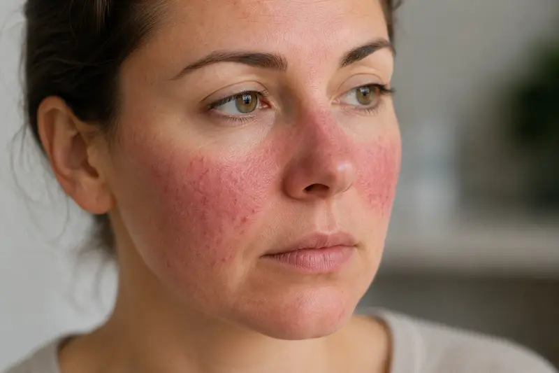
Understanding type 2 rosacea requires understanding two things simultaneously: what the conventional subtype classification system tells you, and what it doesn’t. The four rosacea subtypes describe what the skin looks like. They do not describe what is creating the skin presentation, why it progresses, or how to reverse it. Two patients with identical type 2 rosacea presentations can have completely different upstream drivers — and without identifying those drivers, the label is clinically incomplete as a foundation for treatment.
The Four Types of Rosacea — A Brief Overview
The conventional rosacea classification system identifies four subtypes based on what presents on the skin surface:
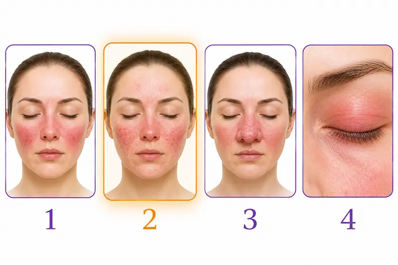
Type 1 — Erythematotelangiectatic Rosacea (ETR)
The vascular-predominant presentation. Characterized by persistent central facial redness, episodic flushing, and visible blood vessels (telangiectasia). No pustules or papules. The skin often has a reactive, sensitive quality that worsens with heat, sun, alcohol, and stress. This is where rosacea most commonly begins — what patients describe as “I just flush easily” or “my cheeks are always red” before the picture becomes more complex.
Type 2 — Papulopustular Rosacea (PPR)
The inflammatory presentation. Characterized by the redness and vascular features of type 1 alongside papules (solid raised bumps) and pustules (pus-filled bumps) that are frequently confused with acne. The critical distinguishing feature: true type 2 rosacea does not produce comedones — no blackheads, no whiteheads. If comedones are present alongside the pustules, the picture is more likely acne or a combination presentation rather than pure rosacea. This subtype represents a meaningful escalation of the inflammatory picture from type 1 — one that reflects a more burdened internal environment, not simply more of the same process.
Type 3 — Phymatous Rosacea
Characterized by skin thickening and tissue overgrowth — most commonly affecting the nose (rhinophyma), producing the enlarged, irregular nasal appearance associated with advanced rosacea. More prevalent in men. Represents chronic inflammatory tissue remodeling rather than the acute inflammatory expression of type 2.
Type 4 — Ocular Rosacea
Eye involvement — watery, irritated, gritty, or light-sensitive eyes, often alongside eyelid inflammation (blepharitis) and meibomian gland dysfunction. Can occur independently of skin rosacea or alongside any of the other subtypes. Frequently unrecognized as rosacea-related because the eye symptoms are managed separately by ophthalmology without connecting them to the skin picture.
These four subtype descriptions are clinically useful as a starting vocabulary. They tell you what the skin is expressing. They do not tell you what is producing that expression, why it progressed from one subtype to another, or what needs to change for the expression to resolve. That is the territory that functional medicine investigates.
What Type 2 Rosacea Is Actually Telling You
The transition from type 1 to type 2 rosacea — from persistent redness and vascular reactivity to the addition of papules and pustules — is not a random escalation. It reflects a specific internal shift: the addition of a hormonal inflammatory layer on top of the vascular dysregulation that was already present.
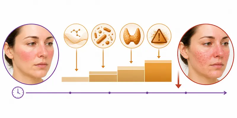
Type 2 rosacea’s pustules are particularly informative. Pustules form when the sebaceous glands are stimulated to overproduce sebum — and the hormone most directly responsible for that stimulation is testosterone. Testosterone drives sebaceous gland activity; when testosterone or its more potent form DHT is elevated relative to the regulatory hormones that balance androgen activity, the result is excess sebum production that creates the environment in which papules and pustules develop. This is why type 2 rosacea so often overlaps with hormonal acne in the clinical picture, and why the rosacea vs acne differential is particularly complex at this subtype — because the same androgen-sebaceous pathway driving acne is contributing to the inflammatory pustule component of type 2 rosacea simultaneously.
But the testosterone-sebaceous connection is never the only driver. It is one expression of a broader hormonal imbalance that is itself the downstream consequence of multiple compounding upstream dysfunctions: metabolic dysfunction driving insulin resistance that amplifies androgen signaling, gut dysbiosis impairing estrogen clearance through the estrobolome and creating relative estrogen dominance that suppresses progesterone, thyroid dysfunction impairing every detoxification and immune regulatory pathway, and toxic burden maintaining the systemic oxidative stress environment in which the entire hormonal and immune picture becomes progressively more difficult to regulate.
The type 2 presentation, in other words, is the skin’s visible expression of a body that has been asked to manage too many unaddressed internal burdens for too long. The vascular reactivity of type 1 was already present. The hormonal and metabolic drivers that produce pustules have been added. The immune regulatory capacity that might have prevented the escalation has been depleted. And each failed treatment course — each antibiotic that temporarily clears the pustules while damaging the gut environment producing them — removes another layer of the body’s compensatory capacity, making the next escalation more likely and the overall picture progressively harder to address.
The Skin Barrier in Type 2 Rosacea
Type 2 rosacea skin shares the barrier compromise of type 1 but carries an additional layer of inflammatory damage from the pustule cycle itself. The acid mantle is impaired — producing the tight, shiny appearance that is frequently misread as oiliness but is in fact accelerated trans-epidermal water loss through a compromised barrier. Ceramide deficiency is almost universally present. Essential fatty acid insufficiency further weakens the lipid matrix of the barrier, reducing its ability to contain moisture and protect the reactive underlying vasculature from environmental triggers.
The absence of comedones — the single most reliable differentiator between type 2 rosacea and acne — is the most important visual feature for both patient self-assessment and practitioner clinical assessment. Type 2 rosacea produces pustules through a different mechanism than acne: not through follicular occlusion and comedone formation, but through inflammatory mast cell and hormonal sebaceous pathways that produce the pustule without the preceding blackhead or whitehead. If you are looking at pustules without any blackheads anywhere on the skin, you are almost certainly looking at rosacea rather than acne, or a predominantly rosacea picture where the hormonal driver is contributing a pustule component.
When acne treatments are applied to type 2 rosacea skin — as they frequently are when the diagnosis is unclear — the results are predictably damaging. Benzoyl peroxide, high-concentration salicylic acid, and aggressive retinol further strip the already-compromised acid mantle and deplete the vitamin A and antioxidant reserves that type 2 rosacea skin depends on for barrier restoration. Each treatment course that temporarily reduces the pustules while worsening the barrier compromise sets the stage for a more reactive and more treatment-resistant presentation in the next cycle.
The Label Tells You What. The Upstream Investigation Tells You Why.
Here is the most important clinical insight about the rosacea subtype classification that conventional dermatology rarely communicates: two patients with identical type 2 rosacea presentations can have completely different upstream drivers producing that presentation.
Patient A with type 2 rosacea may be primarily driven by SIBO producing histamine overload and mast cell hyperreactivity, with the hormonal component being secondary to gut-driven estrogen recirculation from beta-glucuronidase activity.
Patient B with identical-looking type 2 rosacea may be primarily driven by mercury toxicity impairing deiodinase D1 conversion of T4 to T3, with the resulting functional hypothyroidism reducing gut motility, impairing DAO production, and creating the metabolic environment in which androgen signaling is amplified.
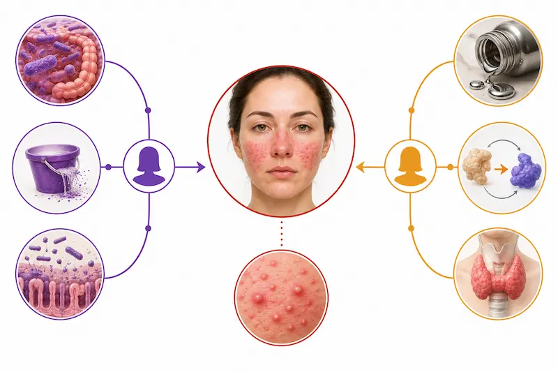
Patient C may have the same surface presentation driven primarily by perimenopause-onset progesterone decline removing the mast cell-stabilizing counterbalance to estrogen, with the HPA axis dysregulation from chronic stress maintaining cortisol elevation that further suppresses thyroid function and amplifies vascular reactivity.
The subtype label is the same for all three. The upstream picture is completely different. And the treatment that addresses one patient’s upstream picture will produce partial results at best for the other two — which explains the profound inconsistency in rosacea treatment outcomes that patients and practitioners alike experience. The label is not specific enough to determine treatment. The upstream investigation is.
Twenty Years of Type 2 Rosacea — Resolved in Five Months
One of my clients had suffered from rosacea for over 20 years. It began as what she described as annoying redness on the cheeks — a classic type 1 presentation that she managed with topical products and accepted as simply her skin’s nature. Over the years, despite repeated and prolonged courses of doxycycline, her rosacea progressed to include pustules alongside the persistent redness — the transition to type 2 that she experienced as a confusing and demoralizing shift. She became uncertain whether she had acne, rosacea, or some combination of both, and the treatments she had been given addressed neither completely.
When she completed her Health History intake questionnaire, the picture that emerged was the picture I see consistently in patients whose rosacea has progressed significantly despite conventional treatment: she had been diagnosed with celiac disease — confirming the gut-immune connection that research consistently documents in rosacea. She had hypothyroidism — impairing every downstream function from gut motility to DAO production to detoxification to immune regulation. She had Raynaud’s syndrome — a vascular autoimmune condition driven by the same sympathetic nervous system and vascular dysregulation pathways involved in rosacea’s own vascular component. She had IBS symptoms, GERD, cramping, and poor sleep — the gastrointestinal comorbidity picture that research confirms is present in virtually all rosacea patients. And she had joint pain, low energy, and low motivation — the systemic burden of two decades of unaddressed upstream dysfunction expressing simultaneously across multiple organ systems.
She had no idea that any of these conditions were connected to her rosacea. She had been managing each one separately, with separate practitioners and separate treatments, and none of the connections between them had ever been made explicit. The celiac disease was managed with a gluten-free diet but without addressing the intestinal permeability and microbiome damage that celiac typically leaves behind.
The hypothyroidism was managed with medication but without investigating the antibody picture, the nutrient cofactors for TPO function, or the toxic burden impairing D1 deiodinase conversion.
The rosacea was managed with doxycycline — which was simultaneously worsening the gut dysbiosis from the celiac, impairing the microbiome diversity that was already compromised by years of gluten-induced intestinal damage, and depleting the B6, zinc, and SCFA-producing bacteria that her DAO enzyme, thyroid function, and immune regulation depended on.
Once we began addressing the upstream picture systematically — gut restoration, thyroid support, nutrient repletion, toxic burden assessment, hormonal rebalancing — her rosacea began responding differently than it had to any previous treatment. Not suppression followed by relapse. Genuine, progressive improvement that continued and deepened as each upstream layer was addressed. By the five-month mark, her rosacea had resolved without medications. Her energy returned. Her joint pain resolved. Her gut symptoms normalized. Her sleep improved. Not because five separate conditions were treated with five separate interventions — but because one upstream environment was addressed, and five downstream expressions of that environment resolved together.
Twenty years of type 2 rosacea. Resolved in five months. Not through a better antibiotic or a stronger topical. Through the upstream investigation that had never happened.
The Label Is a Starting Point. The Investigation Is the Work.
Type 2 rosacea is not a worse version of type 1 that requires stronger treatment. It is a signal that the upstream burden has been accumulating long enough, and without sufficient systemic support, to cross the threshold into a more inflammatory expression. The window for the most complete reversal is earlier rather than later. And as the case above demonstrates, five months of upstream investigation achieved what twenty years of doxycycline could not.
If you have been given a type 2 rosacea diagnosis — or if you have watched your rosacea progress from manageable redness into a more complex picture of pustules, hormonal involvement, and treatment resistance — the most important next step is not a stronger treatment for the label you’ve been given. It is the upstream investigation that the label cannot tell you anything about.
The upstream picture — the gut microbiome, the thyroid status, the hormonal balance, the nutrient sufficiency, the toxic burden — is what determines whether type 2 rosacea continues to progress or begins to resolve. It is measurable. It is addressable. And addressing it is what produces the kind of outcome described above: genuine resolution rather than indefinite management, without medications rather than despite them, in a timeframe that decades of conventional treatment never achieved.
→ Book your Initial Functional Medicine Health and Skin Assessment
P.S. If finances have been the only thing standing between you and starting your functional medicine journey — flexible payment options are available through Cherry on our website. No interest, no impact on your credit score, quick approval, and affordable monthly payments. Apply in minutes on our website.
Q1: How do I know if I have type 2 rosacea?
Type 2 rosacea — papulopustular rosacea — is characterized by the persistent central facial redness and visible blood vessels of type 1 rosacea alongside papules (solid raised bumps) and pustules (pus-filled bumps). The most reliable distinguishing feature is the absence of comedones: type 2 rosacea does not produce blackheads or whiteheads. If pustules are present without any blackheads or whiteheads, the picture is more consistent with rosacea than acne. The skin may appear oily but feel tight and dehydrated — the apparent oiliness reflecting trans-epidermal water loss from a compromised acid mantle rather than genuine sebaceous overactivity. If you are uncertain whether you have type 2 rosacea, acne, or a combination, the comedone rule and the location of lesions (central face for rosacea, lower face and jawline more typical for grade 4 acne) are the most useful starting points.
Q2: Why do I have bumps and pustules with type 2 rosacea?
Type 2 rosacea’s pustules form through a different mechanism than acne pustules. In rosacea, the pustule component reflects hormonal stimulation of sebaceous glands — particularly through testosterone and its more potent form DHT driving excess sebum production — alongside the mast cell activation and immune dysregulation already present in type 1 rosacea. This hormonal-sebaceous driver is itself the downstream consequence of compounding upstream imbalances: insulin resistance amplifying androgen signaling, gut dysbiosis impairing estrogen clearance and creating relative estrogen dominance, thyroid dysfunction impairing detoxification and immune regulatory capacity, and toxic burden maintaining the systemic oxidative stress environment in which the hormonal picture becomes progressively harder to regulate. The pustules are the surface expression of this compounding internal picture — not a separate skin condition superimposed on rosacea.
Q3: Can type 2 rosacea progress to type 3?
Yes — and the progression reflects the same principle as the type 1 to type 2 transition: the upstream burden has continued to accumulate and compound without being addressed, and the skin’s expression has escalated accordingly. Type 3 rosacea (phymatous rosacea) involves chronic inflammatory tissue remodeling — skin thickening and overgrowth — that develops when the sustained inflammatory environment of type 2 rosacea produces fibroblast activation and connective tissue changes over time. This progression is not inevitable. It reflects an internal environment that has been producing sustained inflammation long enough for structural tissue changes to occur. Addressing the upstream drivers that are sustaining the inflammatory environment is the most meaningful intervention available to reduce the risk of progression — earlier in the process rather than after structural changes have already established themselves.
Q4: Can hormones trigger type 2 rosacea?
Yes — and hormonal imbalance is one of the most consistent upstream drivers of the type 2 presentation specifically. Testosterone and DHT stimulate sebaceous gland activity, contributing directly to the pustule component that distinguishes type 2 from type 1. Estrogen dominance — from poor estrogen clearance, gut dysbiosis impairing the estrobolome, or perimenopause-onset progesterone decline — simultaneously stimulates mast cells and drives histamine overload that amplifies the inflammatory picture. Poor progesterone production impairs TPO activity, reducing thyroid hormone synthesis and further compounding every downstream dysfunction. And cortisol elevation from chronic stress or gut-derived physiological stress suppresses thyroid function through TBG elevation while simultaneously activating sympathetic mast cell degranulation. The full hormonal picture — including 24-hour urinary estrogen metabolites, progesterone, cortisol patterns, and androgens — is best assessed through the urinary testing such as Hormone Zoomer rather than standard serum panels.
Q5: Can type 2 rosacea be permanently cured?
The word ‘cure’ implies a one-time intervention that permanently eliminates a condition regardless of what happens subsequently. What functional medicine produces is something clinically more meaningful: resolution of the upstream environment that was producing the type 2 rosacea, alongside the understanding and tools to maintain that environment so that the rosacea does not recur. Patients who address the gut, thyroid, hormonal, nutrient, and toxic burden picture that was driving their type 2 rosacea consistently experience genuine, lasting resolution — not indefinite management of symptoms that return the moment treatment stops. Whether that constitutes a ‘cure’ depends on the definition. What it produces, in clinical practice, is the kind of resolution that decades of conventional treatment consistently failed to achieve.
Q6: Can antibiotics really treat type 2 rosacea effectively?
Antibiotics produce real short-term improvement in type 2 rosacea through their anti-inflammatory properties and antimicrobial action. The clinical reality, however, is that conventional antibiotic therapy for rosacea is usually unsatisfactory because of frequent relapses — because the antibiotics address the downstream expression without touching the upstream gut dysfunction, hormonal imbalance, thyroid insufficiency, and toxic burden driving the type 2 presentation. Each antibiotic course simultaneously damages the gut microbiome that the rosacea picture depends on for immune regulation and DAO activity, further impairing the internal environment. Long-term antibiotic use for type 2 rosacea can also contribute to the gut dysbiosis, estrogen recirculation, and fat-soluble nutrient malabsorption that worsen both the hormonal and inflammatory components of the type 2 picture. For a more detailed explanation of why antibiotics make rosacea harder to treat over time, see our dedicated post on this topic.
Written by Natalie Maibenko – a Certified Functional Medicine Practitioner and Master Esthetician with 22+ years of experience and founder of Unique Verve

Natalie Maibenko
Functional Medicine & Skincare Expert – Helping You Take Control of Your Health and Achieve Lasting Skin Results Nationwide — Virtual Practice
As a Certified Functional Medicine Practitioner my Expertise Encompasses:
- Immune System: frequent illness, UTIs, yeast infections
- Allergies, Asthma
- Skin Problems: acne, cystic acne, rosacea, eczema, dermatitis, ichthyosis, psoriasis, vitiligo, melasma
- Inflammation: arthritis, rhinitis, joint & muscle pain, migraines, headaches
- Sleep Disturbunces, Insomnia
- Gut Problems: IBS/IBD, bloating, acid reflux, gas, constipation, diarrhea, parasites, fungal/yeast overgrowths
- Hormonal Imbalances: PCOS, PMS symptoms, weight problems/inability to lose weight, thyroid problems
- Hair Loss, Alopecia
- Mood Imbalances: anxiety, depression, irritability
- Metabolic Dysfunction, Insulin Resistance, Type 2 Diabetes
- Optimizing Wellness for Successful Pregnancy
- Autoimmune Conditions: Hashimoto’s thyroiditis, grave’s disease, reumatoid arthritis (RA), lupus, etc
- Bone Health: osteopenia/ osteoporosis
- Effective Anti-Aging Strategies without Injectables with the inside-out & outside-in approach
- Detoxification of Heavy Metals, Mycotoxins, Environmental Toxins
- Reversing Breast Implant Illness
- Preparation for the Explant Surgery and Optimization of Wellness & Vitality Post-Explant
